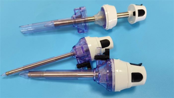

The incidence of incisional hernia after laparoscopic surgery is about 0.1%-3%, which is closely related to the design and operation of the trocar perforators. The risk of abdominal wall tissue injury and poor healing can be significantly reduced by optimizing the trocar structure, material and closure mechanism. The following is a systematic analysis of the design optimization of trocars from the perspective of the causes of incisional hernia.

I. Incisional hernia formation mechanism and perforator correlation
Core causative factors
Fascial defects: The destruction of the rectus abdominis muscle sheath or the integrity of the peritoneum due to trocar puncture holes (especially ≥10 mm) is the anatomical basis of incisional hernia.
Impaired tissue healing: intraoperative high-frequency scalpel thermal injury, postoperative infection, or excessive stretching delay repair of the fascial layer.
Imbalance of abdominal pressure: postoperative coughing and constipation lead to a sudden increase in abdominal pressure and protrusion of the contents of the weak point.
Impact of perforator design
Perforation aperture: the incidence of trocar incisional hernia is 4-6 times higher at 10 mm and above than at 5 mm.
Puncture method: Sharp punctures are more likely to cause fascial tears than blunt separations.
Cannula morphology: Cylindrical cannulas aggravate tissue damage by rotational friction, whereas tapered designs reduce shear forces.
II. Optimization of puncture device design
1. Miniaturization and multi-aperture adaptation design
Reduction of standard aperture: Promote 5 mm as the mainstream puncture aperture (suitable for more than 85% of the procedures), and use a 10 mm cannula only when necessary.
Variable-diameter trocars: Memory alloy or folded structures are used to expand to the desired aperture (e.g., 5→10 mm) intraoperatively and return to the original diameter postoperatively to minimize defects.
Data support: Studies have shown that the incisional hernia rate for 5 mm trocars is only 0.2% compared with 1.8% for 10 mm trocars (Surgical Endoscopy 2021).
2. Innovations in puncture core construction
Blunt detachment technology:
Blunt tip puncture core: replaces the traditional prismatic conical sharp tip and reduces fascial fiber breakage by gradual blunt dilatation (similar to Hasson's technique).
Ultrasound-guided puncture: integrated micro-ultrasound probe in the puncture core, real-time display of the abdominal wall level, avoiding inadvertent puncture of retroperitoneal vessels or organs.
Energy-assisted puncture:
High-frequency electrosurgical coating: Insulation layer plated on the surface of the puncture core, synchronized electrocoagulation for hemostasis during puncture, and reduction of thermal damage diffusion (temperature control ≤70°C).
3. Trocar morphology and surface treatment
Tapered gradient design: the outer diameter of the trocar gradually increases from tip to tail (taper 5°-10°), reducing the tissue shear force during rotational placement.
Anti-adhesion coating:
Hydrophilic coating: reduces frictional resistance between cannula and tissue (40% reduction in coefficient of friction).
Heparinized surface: inhibits fibrin deposition and reduces chronic inflammation caused by postoperative adhesions.
4. Postoperative closure system integration
Pre-positioned fascial suture device:
Sleeve with thread: absorbable suture (e.g., PDS II) is pre-buried in the side wall of the trocar, and the fascial layer is synchronized with the suture when the trocar is removed (the technique of “sewing while retreating”).
Memory alloy closure clip: Nitinol clip is integrated in the end of the trocar, which automatically closes the fascial defect after removal (closure force ≥15N).
Bio-gel closure technology:
The surface of the trocar is loaded with drugs (e.g. fibrin glue), which are released into the puncture channel during extraction to promote tissue adhesion.
5. Optimization of material biocompatibility
Degradable trocars: made of polylactic acid (PLA) or polycaprolactone (PCL), which gradually degrade over 6-12 months, during which time they provide mechanical support and induce tissue regeneration.
Anti-infective coating:
Chlorhexidine/Silver Ion Coating: reduces the rate of postoperative infection (literature shows a 50% reduction in infection-related incisional hernias).
Antibiotic Sustained Release System: sustained release of cephalosporin antibiotics from the carrier cannula for ≥ 72 hours.

III.Clinical Validation and Effectiveness Evaluation
Animal experimental data
Porcine model test showed that the fascial tensile strength of the blunt puncture + pre-suture trocar group was 35% higher than that of the traditional group, and healing was complete at 28 days postoperatively.
Clinical Trial Advances
Biodegradable Cannula: A European multicenter study (n=300) showed that the incisional hernia rate was 0.5% in the PLA cannula group, which was significantly lower than the conventional group at 2.1%.
String closure system: A Korean RCT (n=200) confirmed that the pre-positioned suture technique reduced the incidence of 10 mm incisional hernia from 2.4% to 0.5%.
IV.Future Technological Trends
Intelligent sensing system
Trocar integrated pressure sensors for real-time monitoring of abdominal wall tension and intraoperative indication of risk of overdrawing.
Tissue Engineering Repair
The surface of the trocar is loaded with stem cells or growth factors (e.g. PDGF) to actively promote fascial regeneration.
Magnetic anchored closure technology
Utilizes magnetic materials to achieve sutureless closure and reduce chronic pain due to foreign body residue.
Conclusion
The risk of incisional hernia after laparoscopy can be systematically reduced by miniaturized design, blunt puncture technology, postoperative closure system integration, and biomaterial innovation. In the future, it is necessary to promote the clinical application of the “puncture-operation-closure” integrated solution and optimize the design with long-term follow-up data. Enterprises should focus on the integration of degradable materials and intelligent closure technology to balance minimally invasiveness and safety and reshape the standard process of laparoscopic surgery.
+86 18361958211
marketing@cndonho.com
+86 18361958211
No.2 Zhiwei Road, Qiandeng Town, Kunshan City, Jiangsu Province, China


